MIT Scientists Develop Technique for Better Study of Tissues
| Marco Foronda | | Jan 17, 2015 06:29 AM EST |

(Photo : Fei Chen and Paul Tillberg) Using a new technique that allows them to enlarge brain tissue, MIT scientists created these images of neurons in the hippocampus.
MIT researchers has taken a novel approach to taking high-resolution images using a method that enlarges tissue samples by embedding them in a polymer that swells when water is added.
This technique physically magnifies specimens and images them at a much higher resolution.
Like Us on Facebook
It uses inexpensive, commercially available chemicals and microscopes commonly found in research labs. It should give more scientists access to super-resolution imaging.
The new technique is called "expansion microscopy". It hasn't reached the level of the fine-scale resolution in electron microscopes or the super-resolution microscopes but it won the Nobel Prize last year.
The technique offers an inexpensive way for scientists to examine fine cellular structures at a detailed level using off-the-shelf components.
"Instead of acquiring a new microscope to take images with nanoscale resolution, you can take the images on a regular microscope. You physically make the sample bigger, rather than trying to magnify the rays of light that are emitted by the sample," says senior author of a paper Ed Boyden.
Boyden sees the technology's strength as allowing three-dimensional analysis of tissues. For example, it could reveal the cellular interactions that cause a cancer to spread or reveal a new way to map the brain.
Details of the new research were published in the journal, Science.
Tagshigh-resolution images, expansion microscopy, image enlargement, three-dimensional analysis, tissue, cellular tissues
©2015 Chinatopix All rights reserved. Do not reproduce without permission


EDITOR'S PICKS
-

Did the Trump administration just announce plans for a trade war with ‘hostile’ China and Russia?
-
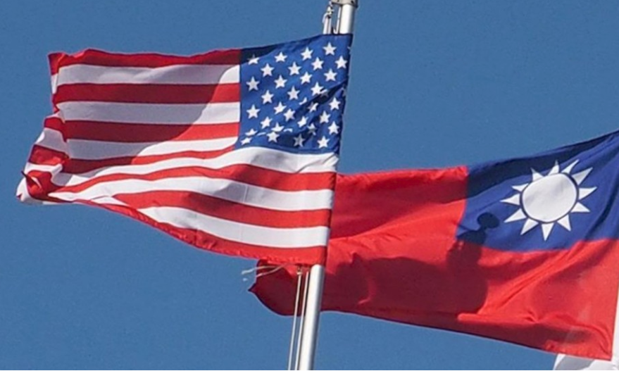
US Senate passes Taiwan travel bill slammed by China
-

As Yan Sihong’s family grieves, here are other Chinese students who went missing abroad. Some have never been found
-
Beijing blasts Western critics who ‘smear China’ with the term sharp power
-

China Envoy Seeks to Defuse Tensions With U.S. as a Trade War Brews
-

Singapore's Deputy PM Provides Bitcoin Vote of Confidence Amid China's Blanket Bans
-

China warns investors over risks in overseas virtual currency trading
-
Chinese government most trustworthy: survey
-

Kashima Antlers On Course For Back-To-Back Titles
MOST POPULAR
LATEST NEWS
Zhou Yongkang: China's Former Security Chief Sentenced to Life in Prison

China's former Chief of the Ministry of Public Security, Zhou Yongkang, has been given a life sentence after he was found guilty of abusing his office, bribery and deliberately ... Full Article


TRENDING STORY

China Pork Prices Expected to Stabilize As The Supplies Recover
-
Elephone P9000 Smartphone is now on Sale on Amazon India
-
There's a Big Chance Cliffhangers Won't Still Be Resolved When Grey's Anatomy Season 13 Returns
-
Supreme Court Ruled on Samsung vs Apple Dispute for Patent Infringement
-
Microsoft Surface Pro 5 Rumors and Release Date: What is the Latest?